Dissertation Research
Our subjective experience of the world is based in our brain’s incredibly complex neural circuitry. The intricate patterns of cell-to-cell connections dictate how sensory information from our environment is transformed into a coherent perception.
The average human brain has around 10 billion neurons and each neuron can make thousands of connections with other neurons, so the circuits formed by these patterns of neuronal connections quickly become unimaginably complex. If we want to get a better idea of how these connections and circuits are organized, we first need to break them down into bite-size chunks and look at each individual circuit component: the synaptic connections formed between only a specific handful of cells. The bulk of my dissertation research in Ed Callaway’s lab at the Salk Institute explored the nature of these connections in greater detail in an effort to better understand how neurons of specific types connect with each other. The project showcased here focused on characterizing the organization of electrochemical connections between neurons—synapses—which form the fundamental basis of information transmission in the brain.
Follow along visually with videos below showing animated slides selected from my dissertation defense (no audio). The core ideas have been translated from TechnicalNeuroJargon to English, displayed as subtitles at the bottom of the slide.
Synapses transform sensory information from our surroundings into a coherent representation of the world. In the part of the brain underlying vision (primary visual cortex or V1), electrical signals arrive from light-sensitive cells in our retinas. These signals, carrying that visual information, travel from neuron to neuron in complex yet precise circuits throughout the brain. At each node in the neural circuit, the information is iteratively processed and transformed; ultimately, this gives rise to our perception of the world (or whatever it is we’re looking at).
One of most striking features of the cortex is its organization into layers, and cells within each layer make robust connections with neurons in other layers. Each layer can be thought of as a computational unit, performing a unique function in transforming visual information. This idea underpins the traditional, long-held (since the ‘80s) notion of an overarching “canonical cortical circuit” motif that governs sensory processing. But this model assumes all neurons in a given layer behave the same way, which we know is not the case. This project therefore asks the question, to what extent do neurons in a specific layer of the cortex communicate with different types of neurons in other layers? By experimentally manipulating the electrical activity in these circuits and seeing how the component neurons respond as a result, we can get a better idea of how these synaptic inputs are organized.
To investigate patterns in these connections between neurons, I used a technique called patch clamp recording. I used tiny glass electrodes to record electrical signals from individual cells in living brain tissue. By comparing signals recorded simultaneously from multiple cells, we were able to determine whether or not those particular cells are connected to each other and part of the same circuit.
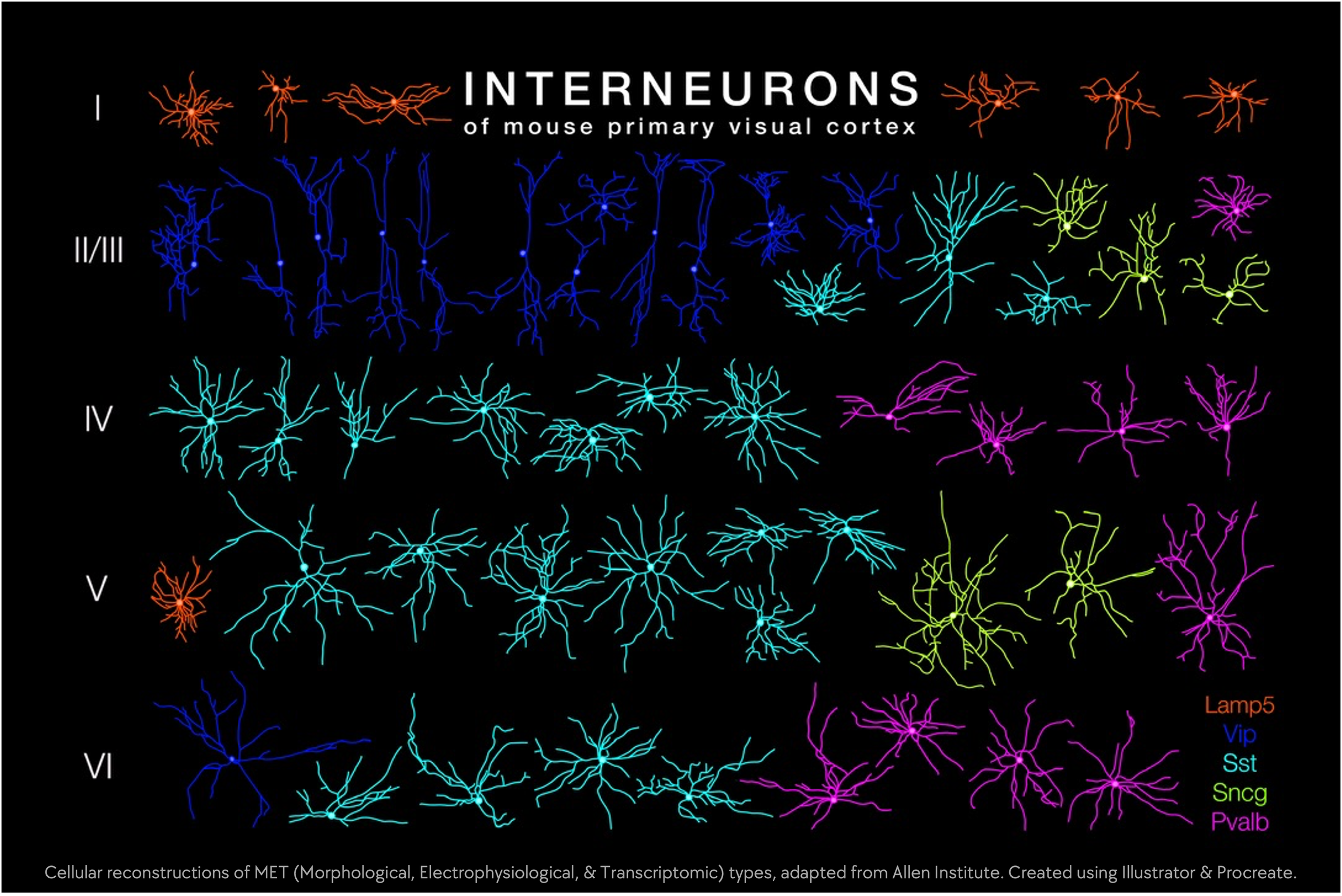
By taking into account other characteristics of each cell, such as what they look like, their precise location, the genes they express, and the types of visual computations they perform, we can get a better understanding of how these cellular connections are organized. By adding up all of the recorded synaptic strengths from each cortical layer (the cells providing the input) to each recorded cell type (the cells receiving the input), we can get a holistic perspective of how much input each layer provides to a specific cell type within a given layer, relative to other layers.
Interestingly, we see that the connectivity strengths are highly dependent on cell type, by output layer, and by recipient layer. In other words, the extent with which cells in one layer connect to cells in another layer depends on the layer of the cell providing the input, the layer of the cell receiving the input, and the subtype of the cell receiving the input. These results ultimately provide us with insight into how visual information is represented within the brain and facilitate discovery of fundamental communication principles between neuronal populations.